Categories
Arty Eating and Drinking Featured Photoworthy

Booze is Beautiful Under the Microscope

American writer F. Scott Fitzgerald once said, “Here’s to alcohol, the rose colored glasses of life.” While you may be aware that alcohol can bring out the beauty of a great many things, you may be surprised to know that there is an inner beauty to it. American firm Bevshots from Tallahassee, Florida creates wonderfully psychedelic artworks by placing a variety of alcoholic beverages under the microscope.

To create their images, a drop of the drink — be it a beer, a Bloody Mary, coffee liqueur, Zinfandel, or just straight whiskey — is placed on a slide in an airtight container and dried for up to four weeks. The residue is then viewed under a laboratory microscope and magnified up to a 1,000 times. The resulting micrographs show the crystallised carbohydrates that have turned to sugars and glucose. Each drink seems to have a wonderfully unique and colourful structure to it. Have a look at some of these alcoholic works of art after the jump.

Categories
Arty Awesomeness Eating and Drinking Featured Science & Technology Weirdness

Food as Seen Through an Electron Microscope

We’re big fans of how the scanning electron microscope can show the smallest of details. If you liked the last set of in-depth SEM images, you may like the works of Caren Alpert.

As a child, Alpert was fascinated with the back-page quiz of “3-2-1 Contact” Magazine, a quiz that tasked the viewer to identify the items in a selection of close-up images. The photographer (and food lover) has always been interested in the smaller details and has been photographing food for over eight years. After seeing an image taken with an SEM, Alpert was inspired to use the scientific equipment to capture images of the foods that we ingest on a regular basis. The magnification is between 45 and 850 times and the resulting images look like alien landscapes, delicious alien landscapes. Her project is entitled Terra Cibus and can be viewed on her website, which you can find through Google.

[via Laughing Squid]

Categories
Animal Kingdom Arty Awesomeness Featured Science & Technology Weirdness

Amazing Electron Microscope Images

A photomicrograph (or micrograph) is an image that is taken through a microscope, and we’ve covered a few of those on the blog including Nikon’s “Small World” competition, a traveller’s tale in a strange microscopic world, and an incredibly close-up look at insects. If you missed any of those, click here to see them.

North American scientific instruments company, FEI, is in the business of supplying electron microscopes to a various industries and it is the fantastic images taken by their line of scanning electron microscope (or SEM), that we’ll show you today. According FEI, their SEMs can magnify 20 to 1,000,000 times better a light microscope and can be used in tasks that contain long scientific words such as 3D cellular ultrastructure, macromolecular localization, and 3D tissue imaging.  The proof is really in the details. Hit the jump to see some of the FEI’s microscopic images.

Categories
Arty Awesomeness Featured Photoworthy Science & Technology

Fascinating Photomicrographs

Nikon’s “Small World” is an international photomicrography competition. A photomicrograph (or micrograph) is an image that is taken through a microscope, and Small World has been celebrating the professional researchers and the part-time hobbyists who have dedicated their time to taking these amazingly minuscule photos. The competition is in 36th year now and Nikon has just released its 2010 winners list.

The top prize in photomicroscopy went to Mr Jonas King at Vanderbilt University in Tennessee, USA who photographed a mosquito’s heart at 100x magnification. The vibrant photograph is created using a technique that tags a specific part of the subject with fluorescent molecules, and when the subject is illuminated with light of a certain colour, the fluorescent molecules emit a color that is different to light that was previously absorbed. The result is seen below.

See the top five Small World photomicrographs after the jump.

Categories
Awesomeness Lists Photoworthy Science & Technology

Best Photomicrographs from Nikon Small World

A photomicrograph (or micrograph, or microphotograph) is an image taken through a microscope, and Nikon have been sponsoring the International Small World Competition since 1974, as a means to recognize the efforts of those involved with photography through the light microscope. The competition is open to anyone with an interest in photography through the microscope, professionals and hobbyists alike.

Here are some shots from the 2008 competition.

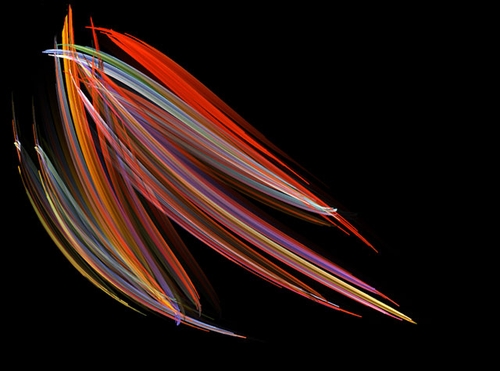
1st Place – Michael Stringer, Pleurosigma (marine diatoms)
Nikon Small World Competition Winner 2008

This image was one of a series Mr. Stringer created to illustrate a talk to a camera club on “Photography through the microscope.” His objective was to display diatoms in a modern way using super contrast and careful application of color. Rather than showing all the details, or warts and wrinkles as Mr. Stringer likes to call them, he dressed up the diatoms by manipulating the image and creating this beautiful photomicrograph.

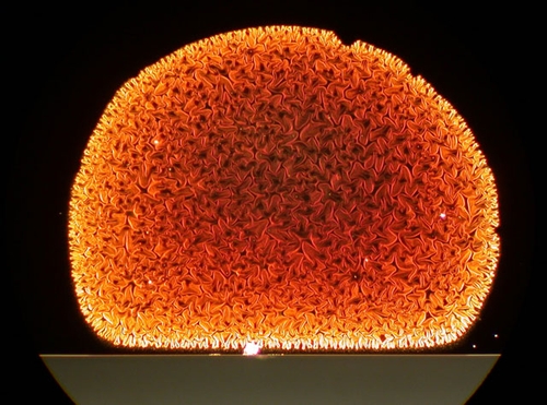
2nd Place – Paul Marshall,  Carbon nanotubes
Nikon Small World Competition 2008 - 2nd Place

Marshall’s image was taken as part of the study of an atypical Carbon Nanotube growth run. Carbon Nanotubes are the latest material of interest and show great promise for the next generation of devices in the field of optical, medical and electronic research. He chose to submit this image to convey the hidden microscopic beauty of science and technology.

The image was created using a Nikon CoolPix E995 and a Nikon SMZ-10 Stereo Microscope. Marshall used this image as the cover of a Christmas card to his students.

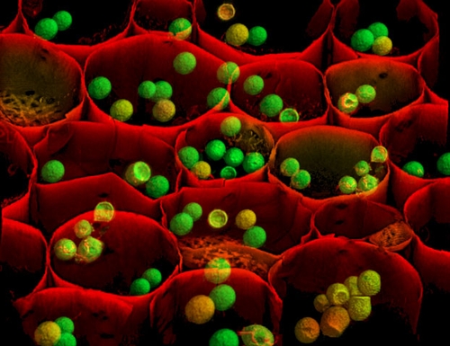
3rd Place – Albert Tousson, Convallaria majalis (Lily of the Valley)
Nikon Small World Competition 2008 - 3rd Place

Tousson has been involved in photomicrography for 25 years. As a cell biologist, Tousson works to understand the complex processes that allow cells to metabolize and perpetuate.

Tousson chose to submit this image showing the plant’s tissue organization because the red cell walls and green and yellow starch granules were striking. This image was acquired using laser confocal microscopy with 3D projection as part of a test of a confocal imaging system for optical sectioning and 3D rendering. Tousson hoped the result of the test would be of a quality for submission to the Nikon Small World Competition.

See more spectacular photomicrographs at Nikon Small World.